|
telescopeѲptics.net
▪
▪
▪
▪
▪▪▪▪
▪
▪
▪
▪
▪
▪
▪
▪
▪ CONTENTS
4.3. Telescope astigmatism
Similarly to coma, astigmatism is an off-axis
point wavefront aberration, caused by obliquity of the incident wavefront relative to
the optical surface. Astigmatism results
simply from
the projectional asymmetry arising from wavefront's inclination to the
optical
surface. The vertical and horizontal
diameter of wavefront's project onto the surface radii that are
effectively different, thus having different focus location for
the tangential plane (the one containing optical axis and
chief ray) and for the plane orthogonal to it
(sagittal plane).
As
illustration below shows,
for an axial light pencil falling onto surface S the surface
radius is of identical value for both tangential and sagittal
cross section, equal to one half of the diameter of a sphere it is a
part of. For an inclined pencil surface radius remains unchanged for
tangential cross section, but the sagittal (horizontal) cross section
projects onto the section of a smaller circle with the diameter
Ri, thus focusing closer
than the vertical section (both cross sections shown are in the same plane).
All vertical sections of the astigmatic
wavefront have common focus, different than the common focus of all
horizontal sections. That constitutes geometry of astigmatism.
Shown is geometry for the stop on the surface; with the chief ray defined
as one passing through the center of aperture stop, it is obvious that
placing stop in front of the surface here would increase, and an internal stop
would decrease astigmatim.
Note that the illustration also reflects other elements of focusing
geometry for abaxial focal points, but what indicates astigmatism
is inequality of surface radii onto which project tangential and
sagittal cross section of the incoming wavefront.
Another peculiarity of astigmatism is that a cross-section along any
horizontal or vertical wavefront diameter (i.e. in the sagittal and
tangential plane)
remains spherical, but with the radius of curvature varying
with the pupil angle. That is what causes the wavefront form as a whole to deviate from
spherical.
For displaced stop -
either first optical surface significantly separated from the aperture
stop, or secondary and tertiary surfaces (whose stop is formed by a
preceding surface) - other surface properties, such as shape, position and conic, also
can influence the size of astigmatism. This is due to the displaced stop
for these
 surfaces changing the surface
radial asymmetry profile. As illustrated at left, the only configuration
that completely corrects for astigmatism is a sphere with the
stop at the center of curvature (1). It is apparent from the two
axes - that of the wavefront and that of the section of the sphere -
coinciding, thus positioning the wavefront symmetrically vs. surface.
Any other stop distance will not be fulfilling this condition,
reintroducing off-axis aberrations (2). For conic forms other
than the sphere, primary astigmatism can be either corrected with
appropriate stop position (closer to mirror with smaller - i.e. negative
- conics), or only minimized (with conics larger than zero). However,
secondary astigmatism, even if low, remains. For a given stop location
other than sphere's center of curvature, the negative conics will
generate less astigmatism than the sphere if their shape results in less
of a surface vs. wavefront positional asymmetry. On the illustration,
that is the case with the stop position 2, as conics toward
parabola and hyperbola open wider than the sphere for given radius of
curvature (however, as the surface section outlined by the wavefront is
not symmetrical about its axis, coma will be generated even if primary
astigmatism is cancelled, with one half of the wavefront flatter than
the other one). surfaces changing the surface
radial asymmetry profile. As illustrated at left, the only configuration
that completely corrects for astigmatism is a sphere with the
stop at the center of curvature (1). It is apparent from the two
axes - that of the wavefront and that of the section of the sphere -
coinciding, thus positioning the wavefront symmetrically vs. surface.
Any other stop distance will not be fulfilling this condition,
reintroducing off-axis aberrations (2). For conic forms other
than the sphere, primary astigmatism can be either corrected with
appropriate stop position (closer to mirror with smaller - i.e. negative
- conics), or only minimized (with conics larger than zero). However,
secondary astigmatism, even if low, remains. For a given stop location
other than sphere's center of curvature, the negative conics will
generate less astigmatism than the sphere if their shape results in less
of a surface vs. wavefront positional asymmetry. On the illustration,
that is the case with the stop position 2, as conics toward
parabola and hyperbola open wider than the sphere for given radius of
curvature (however, as the surface section outlined by the wavefront is
not symmetrical about its axis, coma will be generated even if primary
astigmatism is cancelled, with one half of the wavefront flatter than
the other one).
FIG. 43 illustrates the form of
astigmatic wavefront deformation for primary
astigmatism, and the resulting geometric (ray)
aberration.

FIGURE 43A:
RIGHT: Primary astigmatism
on mirror surface, as a result of the projected diameter of the incoming
wavefront (Wi) varying with the radial orientation. For the
inclination angle shown, the width of vertical, or tangential (with
the tangential plane determined by the axis and chief ray) wavefront
projection onto the surface, given by cosαD,
is at its minimum,
gradually increasing with the radial angle around the chief ray to the
maximum projection width D in the orthogonal
(horizontal, or sagittal) orientation. With the
wavefront sagitta (depth) constant, its radius (the focal length) varies
with the square of diameter. Being of the smallest diameter, the wavefront section
in the tangential (vertical) plane focuses closest, and the
wavefront section
in the sagittal plane farthest away from the mirror.
LEFT: The actual wavefront deviation from the
corresponding reference sphere (red dots): for the reference
sphere
centered at the mid point of defocus M, and those centered at either sagittal or tangential focus (S
and T, respectively; the deviation for the former has
cylindrical form oriented vertically, for the latter horizontally).
All three spheres coincide at the red dotted line. The P-V error is identical at all three focus locations; however, the
deviation averaged over the wavefront (RMS error) is lower at the mid-focus by a
factor 0.82, making it the best (diffraction) focus. Solid blue line in
the best focus wavefront deviation (M) represents the deviation along
the central cross-section of the actual wavefront vs. perfect reference
sphere centered at the mid-focus point. The dashed blue line is a
projection of the deviation along the wavefront edge, indicating
saddle-like shape of the wavefront deformation.
 43B: Ray geometry of astigmatism is not immediately obvious. It is determined by the inequality of the two perpendicular radii, so that all sections of the wavefront parallel to the shorter radius - the vertical one in this illustration - focus in a horizontal line closer to the objective, and all sections of the wavefront parallel to the perpendicular to the former, longer radius, focus into a vertical line farther away from the objective. This ray geometry produces a round ray spot plot midway between the two line foci. Below are the corresponding wavefront deformations for the three axial focal points. Since the reference sphere for F1 is with the corresponding, shorter radius, wavefront deformation in the vertical direction is zero (since the wavefront maps are for the Zernike aberration form, their color code shows zero deviation for the intersection with zero mean, while in the classical form zero deviation is what is coded red for the two cylindrical deviation forms; for the saddle-like, it is identical to the Zernike form, light blue).
Gaussian focus for astigmatic wavefront lies on the
Petzval surface of an optical
surface, or a system. Balancing defocus aberration for this point -
located on the opposite direction from the sagittal focus, and at
identical distance from it as the best focus - is zero, and the
wavefront error is largest. Between the sagittal and tangential focus, ray disturbance resulting from the astigmatic wavefront deformation
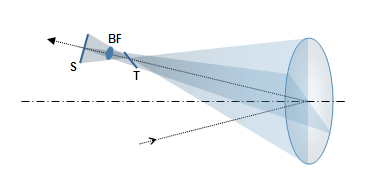
takes on rather peculiar form (FIG. 44).

 FIGURE
44:
Geometry of the astigmatic defocus produced by a mirror with the
stop at the surface: the wavefront radius at the pupil gradually increases
from the minimum in the
tangential (vertical) plane to the maximum in the orthogonal to it
sagittal plane. Consequently, all wavefront meridians focus at a
different length, producing longitudinal defocus, as an axial separation between tangential and sagittal focus. At the
sagittal plane focus S it forms sagittal line, contained in the
tangential plane. And at the tangential plane focus T
it
forms tangential line, laying in the sagittal plane. The lines
transform into ellipses of decreasing eccentricity toward the inside
of defocus zone. Midway between the two lines is the circle
of least confusion (BF), which is the location of best
astigmatic focus. FIGURE
44:
Geometry of the astigmatic defocus produced by a mirror with the
stop at the surface: the wavefront radius at the pupil gradually increases
from the minimum in the
tangential (vertical) plane to the maximum in the orthogonal to it
sagittal plane. Consequently, all wavefront meridians focus at a
different length, producing longitudinal defocus, as an axial separation between tangential and sagittal focus. At the
sagittal plane focus S it forms sagittal line, contained in the
tangential plane. And at the tangential plane focus T
it
forms tangential line, laying in the sagittal plane. The lines
transform into ellipses of decreasing eccentricity toward the inside
of defocus zone. Midway between the two lines is the circle
of least confusion (BF), which is the location of best
astigmatic focus.
Aberration
function for primary astigmatism given in
Table 4 is for tangential focus,
which is identical to that for the sagittal focus, except for the
orientation. Neither coincides with the best (diffraction) focus, which
is located midway between the two, hence requires correction by defocus.
Since for given longitudinal aberration the P-V wavefront error for
primary astigmatism equals that for defocus, the defocus added to it
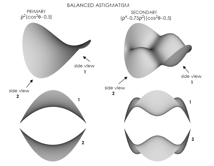
equals half the P-V wavefront error of astigmatism. Aberration function for the
wavefront error of primary
(lower-order) astigmatism at the best
focus is given by:
Wa = Aρ2(cos2θ
- 0.5)
(18)
 with A being the astigmatism
peak aberration coefficient, ρ
the normalized (to 1) height in the pupil, and θ
the pupil angle.
It shows that the
wavefront error peaks for
ρ=1
and cos2θ=1
and 0 (that is, for θ=0,
π/2,
π, 3π/2
and 2π),
which is, every 90 degrees (with the successive peaks being of opposite
sign), and orthogonally to the orientations of the
minimum wavefront deviation, occurring for cosθ=√0.5
(for θ=π/4,
3π/4, 5π/4
and 7π/4). It
clearly outlines saddle-shaped wavefront deviation, as illustrated on
FIG. 43 left. with A being the astigmatism
peak aberration coefficient, ρ
the normalized (to 1) height in the pupil, and θ
the pupil angle.
It shows that the
wavefront error peaks for
ρ=1
and cos2θ=1
and 0 (that is, for θ=0,
π/2,
π, 3π/2
and 2π),
which is, every 90 degrees (with the successive peaks being of opposite
sign), and orthogonally to the orientations of the
minimum wavefront deviation, occurring for cosθ=√0.5
(for θ=π/4,
3π/4, 5π/4
and 7π/4). It
clearly outlines saddle-shaped wavefront deviation, as illustrated on
FIG. 43 left.
Note that the maximum wavefront error given by
Eq. 18 - which gives
±
wavefront deviations, not the peak-to-valley error - is one half of the
peak aberration coefficient, which equals the P-V error. Numerically, it
is identical to the P-V error at either sagittal or tangential focus, Wa=Aρ2cos2θ, but its RMS value is smaller by a factor of 1/√1.5.
When the point of maximum deviation in the tangential
(vertical) plane is closer to the center of reference sphere than its
reference sphere point - i.e. when the tangential focus is precedes
sagittal in the direction of light travel (or, simply put, is closer to
the objective) - the wavefront error of astigmatism is negative.
That is the sign of astigmatism in a concave mirror, illustrated on
FIG. 28. There is no difference in appearance between positive and
negative Seidel astigmatism, since the pattern is merely rotated by
90°, and has an inherent 180-degree rotational symmetry
at the best focus location (FIG. 30). The
peak aberration coefficient A,
which equals the peak-to-valley wavefront error, is given by:
A= aα2d2 = ah2/4F2 (18.1)
with
a being the
astigmatism aberration coefficient (simplified notation for
2w22
in Table 4), h the point height in image
plane,
α the field angle
in radians (with
α=h/f
for object at infinity, f being the focal length) and d
the pupil (aperture) radius. The aberration coefficient a
is, in general form for a single optical surface, given by
a=-0.5n2[(1/n'I)-(1/nO)],
where I, O are the image and
object distance, and n, n' are the refractive index of
incidence and refraction or reflection, respectively.
For a concave mirror and stop at the
surface, n'=-n, I=Of/(O-f), f=R/2, and the coefficient is given by:
aM
= n/R (18.2)
with R being the mirror radius of
curvature. For mirror
in air oriented to the left, n=1 and the aberration
coefficient is aM=1/R.
The sign of aberration coefficient indicates the tangential wavefront
radius shorter than sagittal, and the sagittal line farther away from
the mirror, as shown on FIG. 43-44. Positive astigmatism has this order
reversed. From another perspective, the astigmatism wavefront error is
negative when the optical path difference from the point of peak
deviation in the tangential plane is smaller than the radius of a
perfect reference sphere (the one centered at the mid point between
tangential and sagittal focus).
So, the direct relation for the P-V wavefront error of the primary
astigmatism of a concave mirror can be written as:
Wa =
α2d2/R =
α2D/8F =
h2/8fF2
(18.3)
Shift to the best focus location
is half the longitudinal aberration from either of the two line foci. In
terms of the peak aberration coefficient A, the needed wavefront
error of defocus from either
tangential or sagittal focus to best focus location is
±(A/2).
While the P-V error remains unchanged for all three focus locations -
sagittal, tangential and midway between these two - the latter has the
RMS wavefront error smaller by a factor of 2/√6.
As a result, this focus location has has the highest peak diffraction
intensity, making it the best focus location.
The best focus RMS wavefront error
in terms of the peak aberration coefficient - or P-V wavefront error -
is given by:
ωa
= A/241/2
(19)
Thus the P-V wavefront error of astigmatism corresponding to the
diffraction limited RMS of
ω=1/1801/2
is Wa=(24/180)1/2=0.365,
in units of the wavelength.
It should be noted that for astigmatism, and aberrations in general, the
point of minimum wavefront deviation does not coincide with the point of
PSF maximum, i.e. diffraction focus, when the aberration exceeds certain
level. For aberrations in general, significantly over 1/2 wave P-V.
Specifically for astigmatism, the PSF peak at
 the mid focus becomes
slightly lower than those at the tangential and sagittal foci - despite
the former still retaining lower RMS wavefront error - at 1 wave P-V
(left).
With further P-V error increase, the mid focus PSF peak sinks lower vs.
tangential/sagittal, down to six times lower intensity at 2 waves P-V
(the overall pattern is similar to the longitudinal
PSF peak shift with
spherical aberration, only with significantly larger
maximum-to-mid-focus PSF peak differential). Minimum wavefront error is
always for defocus P-V error half that of astigmatism, i.e. at the mid
focus, but for errors larger than 1 wave P-V PSF peaks in the proximity
of tangential and sagittal focus. PSF peak at the mid focus at 2
waves P-V is identical to that at 3 waves, despite the P-V/RMS error
being 50% larger for the latter. Presence of a larger central
obstruction alters longitudinal intensity distribution in a way that the shift occurs at somewhat less
than 1 wave P-V, with the peak-to-mid-focus differential greater at 1
wave P-V, but smaller at 2 waves. the mid focus becomes
slightly lower than those at the tangential and sagittal foci - despite
the former still retaining lower RMS wavefront error - at 1 wave P-V
(left).
With further P-V error increase, the mid focus PSF peak sinks lower vs.
tangential/sagittal, down to six times lower intensity at 2 waves P-V
(the overall pattern is similar to the longitudinal
PSF peak shift with
spherical aberration, only with significantly larger
maximum-to-mid-focus PSF peak differential). Minimum wavefront error is
always for defocus P-V error half that of astigmatism, i.e. at the mid
focus, but for errors larger than 1 wave P-V PSF peaks in the proximity
of tangential and sagittal focus. PSF peak at the mid focus at 2
waves P-V is identical to that at 3 waves, despite the P-V/RMS error
being 50% larger for the latter. Presence of a larger central
obstruction alters longitudinal intensity distribution in a way that the shift occurs at somewhat less
than 1 wave P-V, with the peak-to-mid-focus differential greater at 1
wave P-V, but smaller at 2 waves.
Note that plots are for negative astigmatism, with the tangential focus
closer to the objective than sagittal.
The form of the aberration coefficient shows that the astigmatism
wavefront error, unlike coma and spherical aberration, doesn't change
with object distance. This is expected consequence of astigmatism of a
mirror - as well as that of a lens (contact) objective - being a result of
the asymmetry of wavefront projection onto the surface, rather than a product of
the wavefront/surface interaction.
Astigmatism
ray aberrations
can also be expressed
in terms of the peak aberration coefficient A as:
L = 8AF2,
T = 4FA
and
Ta = 4A/D
(20)
for the longitudinal, transverse
and angular astigmatism, respectively (the last two as the circle
of least confusion diameter). After substituting for
A and mirror aberration coefficient
a, the mirror transverse aberration - as the circle of least confusion
diameter - can be also expressed as T= -Dα2/2
= -h2/2DF2
for object at infinity, with h being the point height in the image plane
(note that aperture D needs to be in the metric used for the
coefficient calculation, which also becomes the metric of transverse
aberration). Since focal lengths of the astigmatic wavefront do not
change with the height in the pupil (i.e. the zonal height), transverse
astigmatism changes in proportion to the normalized pupil ray
height ρ. With h=αf,
f being the focal length, angular astigmatism Ta=T/f=α2/2F,
where F is the focal ratio number F=f/D.
Longitudinal astigmatism L=-fα2.
There are simple geometric
relationships between the circle of least confusion diameter and the sagittal and
tangential line length, as well as between the three and
the longitudinal aberration. The line length is double the circle
diameter, and the longitudinal aberration is numerically greater than either line by a factor of
F
(the focal ratio), as illustrated in FIG. 44. Hence, the line length equals L/F, and the
geometric blur (circle diameter) L/2F. Since A
expressed in units of a wavelength gives the P-V wavefront error for
that wavelength, the geometric blur at the circle of least confusion
in Airy disc diameters for the same wavelength is given by 4Wa/2.44.
For Wa=0.365, the 0.80 Strehl level, it gives astigmatic
blur of 0.60 Airy disc diameters.
The transverse aberration in
terms of the RMS wavefront error is Ta= ω√384/2.44,
and in terms of the peak-to-valley error Ta=4A/2.44
(for
ω
and A in units of the wavelength), both expressed in
units of the Airy disc diameter. That makes the astigmatic geometric blur significantly
smaller for given amount of wavefront aberration than
geometric blur for
either spherical aberration or coma (FIG. 45). It is a stark
remainder that optical criteria can
not be reduced to geometrical considerations; it is the underlying realm
of electromagnetic field that determines the properties of the point
object image.
 FIGURE
45: The ray spot size (top) and actual diffraction patterns for 0.37 wave
P-V wavefront error of primary astigmatism (resulting in 0.80
Strehl, thus comparable to 1/4 wave P-V of primary spherical
aberration). Perfect diffraction pattern is to the left. Geometric
blur diameter at the best focus location (balanced primary
astigmatism) is only 0.6 Airy disc
diameters. There is no rays outside the Airy disc, yet considerable
amount of energy has spread out from the spurious disc - result of
the complex wave interference around best focus point (not
unexpected, considering that rays focused to a point still
produce a pattern). Compared to
spherical aberration and coma, the energy spread is concentrated
closer to the disc. FIGURE
45: The ray spot size (top) and actual diffraction patterns for 0.37 wave
P-V wavefront error of primary astigmatism (resulting in 0.80
Strehl, thus comparable to 1/4 wave P-V of primary spherical
aberration). Perfect diffraction pattern is to the left. Geometric
blur diameter at the best focus location (balanced primary
astigmatism) is only 0.6 Airy disc
diameters. There is no rays outside the Airy disc, yet considerable
amount of energy has spread out from the spurious disc - result of
the complex wave interference around best focus point (not
unexpected, considering that rays focused to a point still
produce a pattern). Compared to
spherical aberration and coma, the energy spread is concentrated
closer to the disc.
As expected due to its uniformly dense
geometric blur, the smallest RMS blur radius for astigmatism is at the location
of the circle of least confusion. It is given by rRMS=FA√2,
or smaller by a factor of
√0.5
than the radius of the circle of least confusion. In units of the Airy
disc diameter, the RMS blur diameter is RRMS=A√2/1.22,
for the peak aberration coefficient A in units of the wavelength.
In terms of the RMS wavefront error of astigmatism
ωa,
the RMS blur diameter in units of Airy
disc diameter is RRMS=4√3ωa/1.22.
EXAMPLE: A 200mm f/5 concave mirror,
d=100, R=-2000, F=5. Setting θ=0
and ρ=1,
the peak wavefront error at h=1.4mm off-axis, giving the field angle α=1.4/1000=0.0014,
is W=A/2=α2d2/2R=-0.0000049mm.
The P-V wavefront error is
twice greater - equal to the peak aberration coefficient - or
-0.0000098mm. In units of 550nm (0.00055mm) wavelength, it is 0.0178, or 1/56 wave.
Consequently, the RMS wavefront error
ω=A/√24=0.000002mm
or, in units of 550nm wavelength, 1/275 wave. The transverse
astigmatism (circle of least confusion diameter) is
T=4FA=20α2d2/R= =0.000196mm,
or 0.03 Airy disc diameters, and angular astigmatism
Ta=T/f
is 0.000000196 radians, or
206.265x0.000000196=0.04 arc seconds.
Since both, wavefront error and geometric (ray) aberrations are
directly proportional to the aberration coefficient, it implies that
they are in a constant proportion themselves. In other words,
doubling the wavefront error also doubles the geometric aberration.
The RMS blur radius rRMS=FA√2=0.0000098mm,
and the RMS blur diameter in units of the Airy disc diameter is
RRMS=A√2/1.22=0.0207,
for A in units of the wavelength.
Aberration coefficient of
astigmatism doesn't change with object
distance. For relatively close objects,
transverse astigmatism increases as fi/f,
fi
being the image-to-pupil separation. However, it doesn't affect the wavefront
error: since the wavefront radius is also longer by the same ratio, identical nominal wavefront error results in proportionally greater
longitudinal and transverse aberration.
For the aperture stop displaced from mirror
surface, the aberration coefficient of astigmatism changes in
proportion to Δ2[Kσ2+(1-σ)2]/(σR)2,
with σ
being the mirror-to-stop separation (positive in sign) in units of the
mirror radius of curvature. The astigmatism aberration coefficient is:

Needed stop separation for zero astigmatism
is given by σ=[1-√|K|]/(K+1). Thus, astigmatism is canceled for
σ=0.5
with a paraboloid and σ=1
with a sphere. The relation
is not defined for K=-1 (parabola), but implicates σ=0.5 limit for
K approaching -1. For positive values of
the conic K, the aberration coefficient cannot be zero regardless of the
stop position, since both Kσ2
and (1-σ)2
are positive.
Unlike coma, change in astigmatism caused by
displaced aperture stop is independent of object distance.
Aberration coefficient of primary astigmatism for a
lens with the aperture stop at the surface is identical to the one given for
concave mirror (Eq. 19.1), with the radius of curvature R
replaced by 2f (Eq. 99),
f being the lens' focal length. For a contact doublet, it gives the peak
aberration coefficient as a sum of the aberration coefficients at the first
and second lens, respectively, as:

with
α being the field angle, and
f1,f2
the respective lens focal lengths (keep in mind that focal length of a
negative lens is numerically negative in the left-to-right Cartesian coordinate
system). Change of the stop position results in change of the
aberration coefficient only with systems not corrected for spherical
aberration, or coma, or both. Since modern refractor objectives commonly
are aplanats, their astigmatism is not affected by the stop
position. As already mentioned, wavefront error of astigmatism of the
contact doublet doesn't change with object distance.
Strongly curved surfaces give rise to higher order
aberrations, and astigmatism is no exception. Although relatively rare
in amateur telescopes, secondary (higher-order)
astigmatism can be significant in systems with sub-aperture
correctors, as well as in eyepieces. Unlike the P-V wavefront error
of primary astigmatism, which is at either sagittal or tangential focus
described with quadratic function (parabola, blue plot on FIG. 46
left), that of secondary
astigmatism, given as
Wa=Aρ4cos2θ,
is quartic (4th power) function with a single turning point (red plot on
FIG. 46 left). The shape of
wavefront deviation is generally similar to that of primary astigmatism,
but its near-cylindrical form has larger near-flat middle area and more
strongly curved edges. The wavefront shape implies that secondary
astigmatism forms longer sagittal/tangential focus lines when
unbalanced, or balanced with defocus.

FIGURE 46: The form of wavefront deviation of
secondary (higher-order) astigmatism (red) vs. that of primary
astigmatism (blue). For given P-V wavefront error, the secondary
astigmatism deviation, in its pure form (left), is flatter for central
zones, and steeper toward the edges. Balanced with defocus alone, it
takes a form similar to balanced primary astigmatism (shaded gray), only
the saddle is flatter. It is minimized when balanced w/primary
astigmatism, as illustrated with 3D plots below.

Similarly
to that of the primary, the error of secondary astigmatism at either
sagittal or tangential focus (left; tangential focus has the same shape
simply rotated by 90° about vertex) is
reduced by refocusing to the best focus midway between them (right). In
either case, the P-V error remains the same, but the RMS error is
reduced, and smaller for given P-V error for secondary astigmatism at
both focus location.
The more efficient way of minimizing secondary
astigmatism is, however, balancing it with the lower-order form of
opposite sign. The error is minimized with adding the primary
astigmatism
with the peak coefficient smaller by 25%, with Was=A(ρ4-0.75ρ2)(cos2θ-0.5),
implying that the P-V error of of secondary astigmatism balanced with
the lower-order form is smaller by a factor of 0.25 (fourfold) than at either
sagittal or tangential focus. The RMS error is smaller than that of
balanced primary astigmatism of identical P-V error by a factor √0.6.
However, due to the different rates of aberration
increase for the two astigmatism forms (2nd vs. 4th power of field angle
for the primary and secondary form, respectively) this optimized
balancing is only possible at a narrow field zone. Usually, primary
astigmatism is left to be larger initially, with the two forms balancing
farther off axis and secondary astigmatism quickly becoming dominant after that.
◄
4.2. Coma
▐
4.4.Defocus
►
Home
| Comments
|