|
telescopeѲptics.net
▪
▪
▪
▪
▪▪▪▪
▪
▪
▪
▪
▪
▪
▪
▪
▪ CONTENTS
◄
13.6. Eye chromatism
▐
13.8. Eye intensity
response
► 13.7. COMBINED EYE ABERRATIONS, DIFFRACTION, RAYTRACING EYE MODELIn any optical system, the effect of wavefront aberrations - either those induced by the eye itself, by the optical train of a telescope, or externally - is adding to the effect of diffraction in further spreading out the energy making up the point-image. Eye is not an exception. At small pupil sizes, eye diffraction is the main determinant of image quality; at large pupil sizes, that role belongs to eye aberrations, and at intermediate pupil sizes, both diffraction and aberrations are significant factors. Not surprisingly, studies give different indications of the maximum average pupil size at which the eye is still diffraction-limited (0.80 Strehl, or better). The range spans from 1.22mm diameter, or as low as 0.76mm when tilt is excluded (Thibos et al. 2002), to 2.8mm (Howland and Howland, 1977). Prevailing view is that the average eye remains diffraction limited up to about 2mm pupil diameter, after which its exponentially increasing aberrations cause the Strehl to plummet (FIG. 236A). However, the perceived image quality is much less affected than the average nominal Strehl, primarily due to the low effective magnification of the eye (in other words, angular size of either dominant diffraction pattern, or dominant aberrated pattern, remain below detection threshold of the eye).
Size of the geometric aberrated blur (i.e. ray spot size) also varies
with the eye model used.
There is a number of optical models of the
eye, from the simplest one by Emsley (1946), with a single
refracting surface on a water-like
medium (1.333 refractive index), to a complex system proposed by Liou and Brennan in 1997,
incorporating aspheric surfaces and varying refractive indici (the
latter is most closely resembling biological eye, and reproducing its
aberrations). The plot is based on eye model with physical dimensions of
the average eye, constructed to produce its type and level of foveal
aberrations. Specs given in table below (surrounding medium for the
cornea and lens is water; their dispersive properties are obtained by
scaling up those for water approximately corresponding to the change in
e-line refractive index).
As presented previously, both monochromatic and chromatic foveal
aberrations are nominally significant at medium to large eye pupils.
Their combined and separate effects are best illustrated on MTF graph (FIG.
236B). Same goes for monochromatic aberrations - which are usually
the primary concern - and their dependence on the pupil size (FIG.
166C,D). The actual size of diffraction pattern, either nearly
aberration-free (at small pupil sizes) or heavily aberrated (at large
pupil sizes), does not change much going from small to large pupil sizes
- certainly much less than the nominal Strehl, or the RMS wavefront
error value (FIG. 236E).
FIGURE 236: (A) Generalized
scheme of the respective magnitudes of aberration-free
(diffraction-limited) and average real (i.e. aberrated) eye, on axis.
Geometric blur size and RMS wavefront error have nearly identical rate
of change, thus both scales apply to the same plot. With all eye
aberrations included, diffraction dominates for pupil diameters smaller than 2mm, about 1.5mm, and for
larger pupils eye aberrations. At ~2mm pupil diameter, diffraction blur, conventionally represented
by Airy disc or by FWHM, is about 2.4 and 1 arc minute, respectively. For larger
pupils, it is enlarged due to eye aberrations, and for smaller pupils
due to diffraction. With defocus corrected, which applies to the
telescopic eye, eye is diffraction limited for pupils diameters smaller
than ~3mm. Relative magnitude of higher-order aberrations increases
with the pupil size, becoming comparable to the defocus-corrected eye
(i.e. plagued only by primary astigmatism) at pupil diameters over 8mm.
Eye aberrations lower object-to-image
contrast transfer, hence the perceived image quality and resolution
limit. Actual effect on image quality, however, depends not only on the
wavefront error, but also on the retinal image size (magnification). An
aberrated image has to be large enough, angularly, to allow the eye to
detect the aberration. Average eye begins to recognize image shape, as its
size exceeds ~3 arc minutes. Any detail below that size appears
point-like to the eye. Consequently, in order for the eye to discern the
effect of wavefront aberrations in the retinal image - whether an
extended detail or a point-object image - has to be larger than ~3 arc
minutes.
That is why the edge-field coma in an f/6 paraboloid is so
inconspicuous; at 15mm off-axis, with the coma wavefront error at 2.5
waves P-V (0.45 wave RMS), the sagittal blur size produced by a 25mm
eyepiece is still only 3.6 arc minutes on the retina. It takes the blur
size of ~5 arc minutes in diameter for the average eye to clearly
recognize that it is not point-like; in this case, it would take ~f/5 paraboloid.
Considering that the astigmatism of standard eyepieces usually adds
significantly to the blur size that far off axis, 5 arc minutes blur
size is probably still borderline between inconspicuous and obvious
level of aberrations for the average observer.
The required minimum spot size for shape
recognition is greater for less contrasty details, as well as for those
that fall in the low-sensitivity range of eye photoreceptor cells, cones
and rods.
A glance at FIG. 236E reveals that despite its very high nominal
aberrations, the aberrated diffraction blur of the average eye is still
significantly smaller than 5 arc minutes for pupils smaller than about
5mm in diameter, thus with relatively small effect on the perceived
image quality. The exception is eye defocus error (assumed corrected in
the patterns shown), which is typically by far the largest eye
aberration. It often very noticeably degrades quality of the visual
image; fortunately, it does not affect the telescope user.
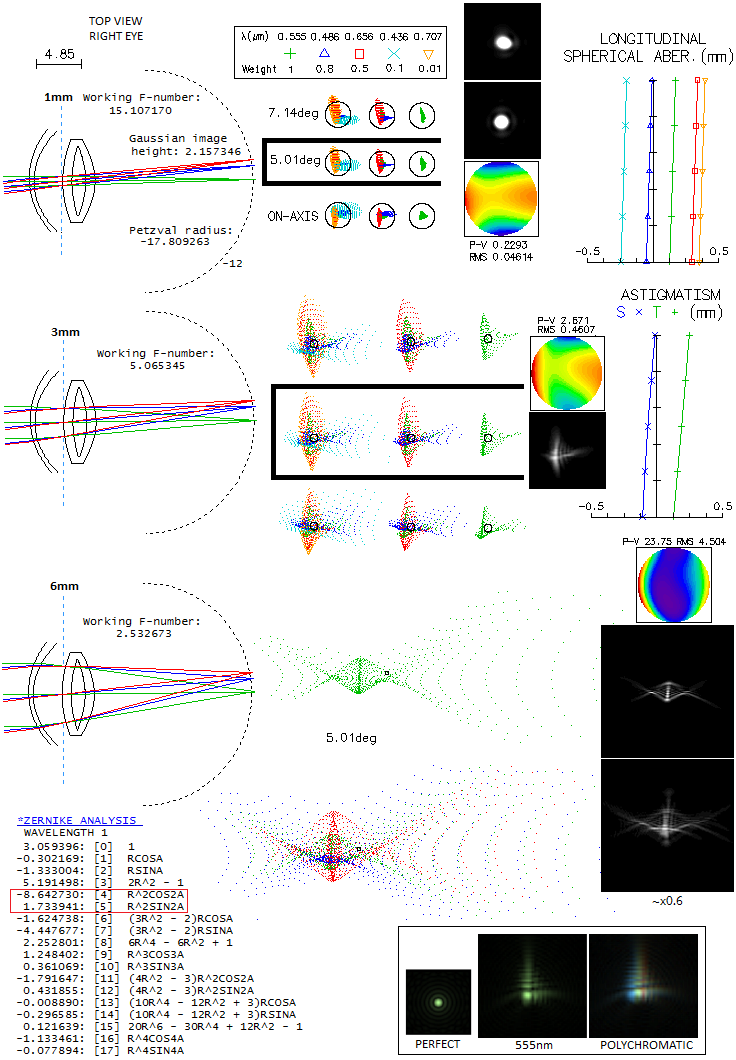
Eye model used here is somewhat different than the one specified above,
main difference being in the type of surface misalignment chosen to
produce asymmetrical blurs, more alike those of the real eye, with the
aberrations magnitude within the average level. The
starting point was model from Chen et al. (Development of a human eye
model incorporated with intraocular scattering for visual performance
assessment, 2012), simplifying the crystaline lens to only two layers, and
limiting misalingment tableo only two surfaces. The radii are modified
to produce 23mm physical focal length. Due to image being formed in a
watery environment with the refractive index n~1.33, the effective focal
length is 17.3mm (this is because the waves get compressed, acting as
shorter wavelengths by the 1/1.33 ratio, hence forming correspondingly
smaller diffraction image; the entire image scales with it). Geometrically,
the focal length is determined by the point where cone converging to the
focus extended toward the opposite end intersects marginal rays of the
pencil entering the cornea. That point is generally slightly inside the eye,
thus the focal lenght is a bit shorter than the phisical separation
between front surface of the cornea and focus (in this case 24.6mm).
The corresponding focal ratio is approximated by f/P, P being the
eye pupil diameter and f the focal length, but its exact
value is given as the ratio of a diameter of the pencil entering cornea
vs. focal length, in general 10-15% smaller. The Petzval radius for
this model is -17.8mm, with the radius of a surface approximating
retinal wall -12mm. The five wavelengths spanning the visual spectrum
are roughly weighted for empirically indicated mesopic sensitivity.
Defocus is omitted as it is not a factor for telescopic eye.
Model is raytraced for three pupil sizes, 1, 3 and 6mm diameter, from
top to bottom, with the corresponding pencil diameters at the cornea
of 1.14, 3.4 and 6.8mm. The field height of 7.14° is chosen so
that the 0.7 field is 5°; this is the angle within 4-8° range,
between optical and visual axis of the eye, due to the fovea being
that much off the optical axis. Hence, the point that represents image
in the center of fovea is that at 5° field (with the 7° is
also being within the range). Longitudinal aberrations for axial chromatism
and astigmatism
don't change with the pupil size. Longitudinal spherical aberration
changes (as wavefront error) in proportion to the 4th power of pupil diameter.
At the 1mm pupil, spherical aberration is entirely negligible. Dominant
aberration is astigmatism, with the
longitudinal axial astigmatism of 0.25mm (nearly 1 diopter) implying
0.24 wave P-V, for 555nm wavelength (from LA/8F2, LA being the longitudinal aberration
and F the focal ratio). Longitudinal chromatism has the form of
primary spectrum, with shorter wavelengths focusing closer than longer ones.
The error for each wavelength, as P-V wavefront error of defocus, is given with
the same formula as for astigmatism, with the only difference being in
the wavelength it is measured against. The blue F and red C line have 0.18mm
of defocus each, translating to 0.2 and 0.15 wave P-V, respectively.
At 0.37mm, the violet g-line comes to 0.47, and red r-line at 0.23mm to
0.18 wave. Diffraction simulation on the bottom shows 555nm wavelenght,
and above is the 5-wavelength simulation - closer to what we actually see -
showing some deformation due to to the blue/violet being shifted toward their
line focus.
At 3mm pupil, f-ratio drops to f/5.1, and both axial astigmatism and
chromatism are larger in inverse proportion to the square of it. The
eye is already far from "diffraction limited" (0.80 Strehl, i.e. 0.0745
wave RMS). Diffraction pattern (shown for the 5 wavelengths) is up to several Airy discs in diameter,
deformed in a more or less asymmetrical blur. At 6mm pupil the focal
ratio is only f/2.5 and the blur yet larger - much more vs. Airy disc than
vs. blur at 3mm pupil. Diffraction patterns are shown somewhat reduced
in size, for 555nm (top) and the 5-wavelengths (bottom). Zernike analysis
shows that the primary aberrations are still dominant (not necessarily
the case with every individual eye), with astigmatism (4,5) still dominant.
Coma (6,7) is roughly half, and spherical aberration (8) one fourth as large (not
to forget that these values add up squared, thus coma is actually only
one fourth, and spherical aberration one 16th of the astigmatism magnitude).
Zernike aberration term value for primary spherical aberration
determines the RMS wavefront error when
divided by 50.5. It gives 1 wave RMS error, i.e. 3.35 wave P-V
(spherical aberration becomes obvious on the wavefront map at the
minimum RMS error focus, but diminishes to negligible at this focus).
That allows to estimate spherical aberration at other pupil diameters,
having in mind that it is just a Zernike term, not the actual aberration.
For 1mm pupil, it would be smaller by a factor 64, or
nearly 1/400 wave P-V. While this is unlikely to be accurate, it is
safe to assume that it is at that pupil size entirely negligible.
Most of the defocus term (3) comes from manually defocusing from the
lowest RMS error focus (3.5 wave) to the focus with the highest energy
encircled within Airy disc, and wider. Best focus in this respect (shown)
encircles 39% energy within 0.01mm radius, while the lowest RMS-error focus,
despite over 20% lower nominal error, only 29%. This illustrates how
unreliable the RMS error - together with the Strehl ratio - becomes
at large magnitude errors. Emerging higher-order aberrations are
trefoil (9,10), secondary astigmatism (11,12) and quadrafoil (16,17).
Diffraction patterns in the box bottom right are from a different eye
model. They show one possible blur shape at a mid-size pupil for 555nm
wavelength, and for the 5 wavelengths specified above. The colors are
there, but brain processing filters them out, and we see a nearly uniform
hue over the polychromatic image (on the left is the unaberrated pattern).
In addition to eye aberrations, other important factors determining
properties of the image formed by the eye are its spectral response,
intensity response, and contrast sensitivity. That comes next.
◄
13.6. Eye chromatism
▐
13.8. Eye intensity response
► |
||||||||||||||||||||||||||||||||

